Pain in bone metastases: from physiopathology to therapeutic options
Il dolore nelle metastasi ossee: dalla fisiopatologia
alle opzioni terapeutiche
Short Review
Pathos 2019, 26; 1. Online 2019, Jan 31
_______________________________________________________________________________________________________
Francesco Amato,1 Erminia Gilda Morrone 2
1 Direttore UOC Terapia del Dolore e CP
AO Cosenza, Centro Hub Regionale
AO Cosenza, Centro Hub Regionale
2 Biologa, Associazione Centro Studi Terapia del Dolore, Cosenza
________________________________________________________________________________________________________
Summary
Cancer-induced bone pain is the most common complication in patients with bone metastases. It causes a significant reduction in patient quality of life. Recent studies using preclinical models have demonstrated the role of the bone marrow microenvironment (osteoclasts, osteoblasts, macrophages, mast cells, mesenchymal stem cells and fibroblasts) in bone pain metastases development. Available analgesic treatments for bone pain metastases, such as opioids that target the central nervous system, come with severe side effects as well as the risk of abuse and addiction. Therefore, alternative treatments are needed to develop more effective and safer targeted therapies.
Riassunto
Il dolore osseo indotto dal cancro è la complicanza più comune nei pazienti con metastasi ossee e provoca una significativa riduzione della qualità di vita del paziente. Recenti studi che utilizzano modelli preclinici hanno dimostrato il ruolo del microambiente del midollo osseo (osteoclasti, osteoblasti, macrofagi, mastociti, cellule staminali mesenchimali e fibroblasti) nello sviluppo del dolore indotto da metastasi ossee. I trattamenti analgesici disponibili per il dolore indotto da metastasi ossee, come gli oppiacei, che interagiscono con il sistema nervoso centrale, hanno gravi effetti collaterali oltre al rischio di abuso e dipendenza. Pertanto, sono necessari trattamenti alternativi per sviluppare terapie mirate più efficaci e più sicure.
Key words
Cancer-induced bone pain, bone marrow microenvironment, bone metastases, osteoclasts, macrophages, radiofrequency ablation
Parole chiave
Dolore osseo indotto dal cancro, microambiente del midollo osseo, metastasi ossee, osteoclasti, macrofagi, ablazione con radiofrequenza
Introduction
Bone homeostasis is characterized by constant remodeling: bone is removed from osteoclasts and reconstructed by osteoblasts. This constant regeneration indicates that the bone tissue is always healthy and fully functioning.
Latest studies on preclinical models studies showedwn how bone marrow microenvironment is involved in metastasis-induced pain, reducing the patient's quality of life.
In osteolytic bone metastases, osteoclast maturation is observed. Osteoclasts increase osteolytic activity destroying microarchitecture (with an increased risk of fractures) and, sometimes, we assist to an increase of hypercalcaemia, while osteoblasts appear to repair tumor cells and protect them from chemotherapy.1
Cross-talk between bone microenvironment and metastasis determines a degradation of the tissue, but also a new arrangement of nerve fibers. The periosteum is densely innervated by myelinated and unmyelinated sensory fibers and by sympathetic fibers that are not normally located close to each other; however, it has been demonstrated how tumor cells induce a reorganization, increasing the density of fibers2 with the formation of structures such as sprouting and neuroma, contributing to the spontaneous generation of episodes of breakthrough cancer pain. Sensory and sympathetic fibers’ reorganization is associated with the increased release of NGF (nerve growth factor). By binding to its TrkA receptor (tyrosinekinase receptor type 1), it can form a compound that, migrating with a retrograde pattern from the periphery to the cell body located at the dorsal ganglion (DRG), stimulates synthesis of neurotransmitters (P substance - calcitonin gene peptide), expression of receptors or channels (bradykinin - P2X3 Purinergic Receptors - TRPV1, transient receptor potential vanilloid 1 - sodium channels) and transcription factors (ATF-3, activating transcription factor 3). In particular, the NGF performs a pronocicective action through pathways that modulate expression and traffic both of sodium channels (Nav) 1.8 and TRPV1 channels, sensitizing them.2-4
Regarding the progression of bone metastases, cells that compose them (and that come from primary tumors such as breast cancer, lung cancer, kidney cancer, sarcomas and prostate cancer) have osteolytic capacity and stimulate osteoblasts.5 In fact, metastatic cells overexpress some factors, among them endothelin 1 (ET1) which, by binding to endothelin A receptors (ETAR) or endothelin B receptors (ETBR) present on osteoblasts, induce their mitogenesis.6
Osteoblasts release the RANKL (nuclear factor kappaB activator) which interacts with the RANK receptor expressed on the osteoclasts. This interaction induces an osteolytic activity increase that enhances bone reabsorption.
The bone matrix reabsorption causes TGFβ (transforming growth factor beta) and IGF-1 (insulin growth factor) release, which enhance the metastatic progression determining a "vicious circle".7-10
During reabsorption process, the extracellular space is characterized by an acidic pH (4.0-4.5) following protons and chlorine ions release expelled by membrane transporters (V-type H + ATPase). Ph decrease sensitizes sensory receptors, in particular TRPV1 ones expressed on nerve fibers, making them more responsive to mechanical, thermal and chemical stress.11,12
The presence of TGFβ (transforming growth factor) and IGF-1 (following the reabsorption of the bone matrix) determines a further up-regulation and sensitization of the TRPV1 along the nerve fibers.13,14
Furthermore, it has been observed that tumor cells secrete adenosine triphosphate (ATP), which has the ability to stimulate osteoclasts by P2X3 receptors.15,16 It is important to observe how tumor cells under hypoxia (present in the bone tissue), by, generating lactate, induce a lowering of the pH, called Warburg effect.17 The phenomenon stimulates the stromal cells to produce cytokines such as IL-6 and IL-8, inducing the formation of NGF and BDNF factors.
By binding to the respective surface receptors receptorkinase A (TrkA) and tropomyosinkinase B (TrKβ) present on the macrophages outer membrane, they activate the release of TNF-alpha and of factors that regulate the inflammation processes such as prostaglandins (PGE2), sensitizing the peripheral nerve ends.18-21
Spinal metastases are localized lesions and represent the most common form of metastasis for many types of solid tumors (40% of patients), particularly lung, breast and prostate. Spinal metastases are usually found at the thoracic level (70%); less frequently at the lumbar (20%) and cervical (10%) levels. More than 50% of patients with vertebral metastases have more than one metastatic lesion.
Spinal metastases are associated with intense pain, pathological fractures, hypercalcaemia and spinal cord compression, which occur in about 20% of patients and lead to urinary and intestinal incontinence and limb weakness. Spinal metastases are a negative prognostic factor (median survival of about 10 months).22
Principles of treatment
Adequate pain management in bone metastases requires a multidisciplinary approach.
Opioids induce analgesia by activating the inhibitory pathway of the periaqueductal grey area (PAG) and the ventromedial rostral marrow. The inhibition of GABA increases the activity of the descending pathways of raphe and reticular nucleus with an increase in serotonin secretion and inhibition of lamina interneurons; the phenomenon consequently induces a suppression of spinal dorsal horn pain impulses transmission.
Anti-inflammatory drugs (NSAIDs) can also be considered: due to their anti-inflammatory and analgesic effect, interacting on the activity of cyclooxygenases, particularly on COX-2 expressed by inflammatory cells, they decrease concentration of prostaglandins and leukotrienes, capable of phosphorylating TRPV1 following the PKA pathway;23-25 obviously, the possible (sometimes severe) side effects induced by these molecules must always be monitored.23-25
Tanezumab is a potential experimental treatment; it is an humanised monoclonal antibody known as a nerve growth factor inhibitor (monoclonal anti-NGF antibody). In preclinical studies on guinea pigs, tanezumab appears to attenuate osteogenic metastases pain in patients with breast cancer and/or prostate cancer; moreover, it has been successfully tested in patients with osteoarthritis, although further clinical studies are required for this indication.26
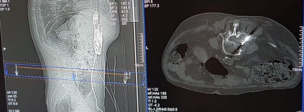
Targeted radiofrequency ablation system is another bone metastases treatment, in addition to radiotherapy. The technique exploits radiofrequency energy emitted through a directional instrument (bipolar electrode) with consequent thermal necrosis of the adjacent tissue and decrease of pain (Figure 1).27
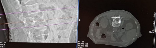
The tumor ablation system by targeted radiofrequency (t-RFA) is used for spinal metastases treatment. A single treatment allows both to locally destroy the tumor and to relieve pain. It also provides the possibility to create site-specific ablation zones and to monitor in real time the area to be ablated within the vertebral body. It is compatible with other systemic therapies and is generally indicated for vertebral metastases treatment in patients undergoing vertebroplasty (Figure 2) (Figure 3).
The directional ablative catheter can be repositioned several times to allow the complete ablation of the metastatic lesion.
This treatment is palliative and its primary objectives are the optimization ofpain control, preservation or restoration of neurological and mobility functions, maintenance of vertebral stability, local tumor control and quality of life improvement.28,29
The mechanisms for succesful pain management in bone metastases include:
1) first of all, the reduction of the tumor mass which decompresses nerve bundles;
2) the destruction of cancer cells that secrete cytokines and growth factors;30
3) the destruction of macrophages that secrete TNF alpha, IL-6, IL-8;30,31
4) the denaturation of cytokines, NGF and BDNF;32
5) destruction of sensory nerve fibers;32-34
Conclusion
After a correct pain assessment and evaluation of pain symptoms, a multidisciplinary approach and an appropriate evaluation of procedures and concomitant pharmacological therapies are all necessary elements, both to ensure the best therapeutic pathway and the correct management of pain, allowing to improve the quality of life of patients with bone metastases, guaranteeing the best possible therapeutic pathway.
Conflict of interest
The authors certify the study was conducted without conflicts of interest.
Published
31th January 2019
Bibliografia
1) Hanqiu Zheng, Yangjin Bae, Sabine Kasimir-Bauer, Rebecca Tang et al. Therapeutic antibody targeting tumor-and osteoblastic niche-derived jagged 1 sensitizes bone metastasis to chemotherapy. Cancer Cell 2017; 32; 6: 731-747. doi: 10.1016/j.ccell.2017.11.002
2) Falk S, Bannister K, Dickenson AH. Cancer pain physiology. British Journal of Pain 2014; 8 (4): 154-162. doi: 10.1177/2049463714545136
3) Sun H. Park, Matthew R. Eber, D. Brooke Widner, Yusuke Shiozawa. Role of the Bone Microenvironment in the Development of Painful Complications of Skeletal Metastases. Cancers 2018; 10(5): 141. doi: 10.3390/cancers10050141
4) Lozano-Ondoua AN, Symons-Liguori AM, Vanderah TW. Cancer-induced bone pain: mechanisms and models. Neurosci Lett 2013; 557: 52–59. doi: 10.1016/j.neulet.2013.08.003
5) Shiozawa Y, Eber MR, Berry JE, Taichman RS. Bone marrow as a metastatic niche for disseminated tumor cells from solid tumors. Bonekey Rep 2015; 4: 689. doi: 10.1038/bonekey.2015.57
6) Kitano Y, Kurihara H, Kurihara Y, Maemura K, Ryo Y, Yazaki Y, Harii K. Gene expression of bone matrix proteins and endothelin receptors in endothelin-1-deficient mice revealed by in situ hybridization. J Bone Miner Res 1998; 13(2): 237–244. doi: 10.1359/jbmr.1998.13.2.237
7) Zheng Y, Zhou H, Dunstan CR, Sutherland RL, Seibel MJ. The role of the bone microenvironment in skeletal metastasis. J Bone Oncol 2012; 2(1): 47–57. doi: 10.1016/j.jbo.2012.11.002
8) Buenrostro D, Park SI, Sterling JA. Dissecting the role of bone marrow stromal cells on bone metastases. BioMed Res Int 2014; 2014: 875305. doi: 10.1155/2014/875305
9) Chirgwin JM, Guise TA. Molecular mechanisms of tumor-bone interactions in osteolytic metastases. Crit Rev Eukaryot Gene Expr 2000; 10(2): 159–178. doi: 10.1615/CritRevEukarGeneExpr.v10.i2.50
10) Guise TA, Kozlow WM, Heras-Herzig A, Padalecki SS, Yin JJ, Chirgwin JM. Molecular mechanisms of breast cancer metastases to bone. Clin Breast Cancer 2005; 5(Suppl. 2): S46–S53. doi: 10.3816/CBC.2005.s.004
11) Lingueglia E. Acid-sensing ion channels in sensory perception. J Biol Chem 2007; 282(24) :17325–17329. doi: 10.1074/jbc.R700011200
12) Holzer P. Acid-sensitive ion channels and receptors. Handb Exp Pharmacol 2009;(194): 283–332. doi: 10.1007/978-3-540-79090-7_9
13) Li Y, Cai J, Han Y, Xiao X, Meng XL, Su L, Liu FY, Xing GG, Wan Y. Enhanced function of TRPV1 via up-regulation by insulin-like growth factor-1 in a rat model of bone cancer pain. Eur J Pain 2014; 18(6): 774–784. doi: 10.1002/j.1532-2149.2013.00420.x
14) Xu Q, Zhang XM, Duan KZ, Gu XY, Han M, Liu BL, Zhao ZQ, Zhang YQ. Peripheral TGF-beta1 signaling is a critical event in bone cancer-induced hyperalgesia in rodents. J Neurosci 2013; 33(49): 19099–19111. doi: 10.1523/JNEUROSCI.4852-12.2013
15) Hoebertz A, Townsend-Nicholson A, Glass R, Burnstock G, Arnett TR. Expression of P2 receptors in bone and cultured bone cells. Bone 2000; 27(4): 503–510. doi: 10.1016/S8756-3282(00)00351-3
16) Morrison MS, Turin L, King BF, Burnstock G, Arnett TR. ATP is a potent stimulator of the activation and formation of rodent osteoclasts. J Physiol 1998; 511(Pt 2): 495–500. doi: 10.1111/j.1469-7793.1998.495bh.x
17) Vander Heiden MG, Cantley LC, Thompson CB. Understanding the Warburg effect: the metabolic requirements of cell proliferation. Science 2009; 324(5930): 1029–1033. doi: 10.1126/science.1160809
18) Zhang XC, Kainz V, Burstein R, Levy D. Tumor necrosis factor-alpha induces sensitization of meningeal nociceptors mediated via local COX and p38 MAP kinase actions. Pain 2011; 152(1): 140–149. doi: 10.1016/j.pain.2010.10.002
19) Binshtok AM, Wang H, Zimmermann K, Amaya F, Vardeh D, Shi L, Brenner GJ, Ji RR, Bean BP, Woolf CJ, Samad TA. Nociceptors are interleukin-1beta sensors. J Neurosci 2008; 28(52):14062– 14073. doi: 10.1523/JNEUROSCI.3795-08.2008
20) Rankin EB, Giaccia AJ, Schipani E. A central role for hypoxic signaling in cartilage, bone, and hematopoiesis. Curr Osteoporos Rep 2011; 9(2):46–52. doi: 10.1007/s11914-011-0047-2
21) Di Pompo G, Lemma S, Canti L, Rucci N, Ponzetti M, Errani C, Donati DM, Russell S, Gillies R, Chano T et al. Intratumoral acidosis fosters cancer-induced bone pain through the activation of the mesenchymal tumor-associated stroma in bone metastasis from breast carcinoma. Oncotarget 2017; 8(33): 54478–54496. doi: 10.18632/oncotarget.17091
22) ASERNIP-S 2017 – Health Policy Advisory Committee on Technology. ASERNIP-S. Technology Brief Update. STARTM Tumor Ablation System. March 2017
25) Raffaeli W, Indovina P. Low dose naltrexone to prevent intolerable morphine adverse events: a forgotten remedy for a neglected, global clinical need. Pain Medicine 2015; 16(6): 1239-42. doi: 10.1111/pme.12704
26) Sopata M, Katz N, Carey W, Smith MD, Keller D, Verburg KM, West CR, Wolfram G, Brown MT. Efficacy and safety of tanezumab in the treatment of pain from bone metastases. Pain 2015; 156(9): 1703–1713. doi: 10.1097/j.pain.0000000000000211
27) Chen F, Tian G, Kong D, Zhong L, Jiang T.Radiofrequency ablation for treatment of benign thyroid nodules: A PRISMA-compliant systematic review and meta-analysis of outcomes. Medicine (Baltimore) 2016; 95(34): e4659. doi: 10.1097/MD0000000000004659
28) Barzilai O, Laufer I, Yamada Y, Higginson DS, Schmitt AM, Lis E, Bilsky MH. Integrating Evidence-Based Medicine for Treatment of Spinal Metastases Into a Decision Framework: Neurologic, Oncologic, Mechanicals Stability, and Systemic Disease. J Clin Oncol 2017; 35(21): 2419-2427. doi: 10.1200/JCO.2017.72.7362.
31) Zhang XC, Kainz V, Burstein R, Levy D. Tumor necrosis factor-alpha induces sensitization of meningeal nociceptors mediated via local COX and P38 map kinase actions. Pain 2011; 152(1): 140–149. doi: 10.1016/j.pain.2010.10.002.
32) Binshtok AM, Wang H, Zimmermann K, Amaya F, Vardeh D, Shi L, Brenner GJ, Ji RR, Bean BP, Woolf CJ, Samad TA. Nociceptors are interleukin-1beta sensors. J Neurosci 2008; 28(52):14062– 14073. doi: 10.1523/JNEUROSCI.3795-08.2008.